Talk to Orthopedic Surgeon on Leg Pain Problem
Asking For Self, Male, 26 Years old, jail road
Homeopathic Dr. Basit Chawla (Psychologist) - Homeopath
DHMS | Karachi
Thank you for your question. In homeopathy, medicine is selected according to the complete symptoms and patient constitution. For accurate remedy selection, a detailed case history is required. You may consult for proper homeopathic treatment.
yes calsium ki kmi lg rhe ha
Dr. Syed Safdar Raza - Orthopedic Surgeon
MBBS ( KE), FRCS (UK), MSc Orthopedic ( London) | Lahore
399 Positive Reviews
Thank you
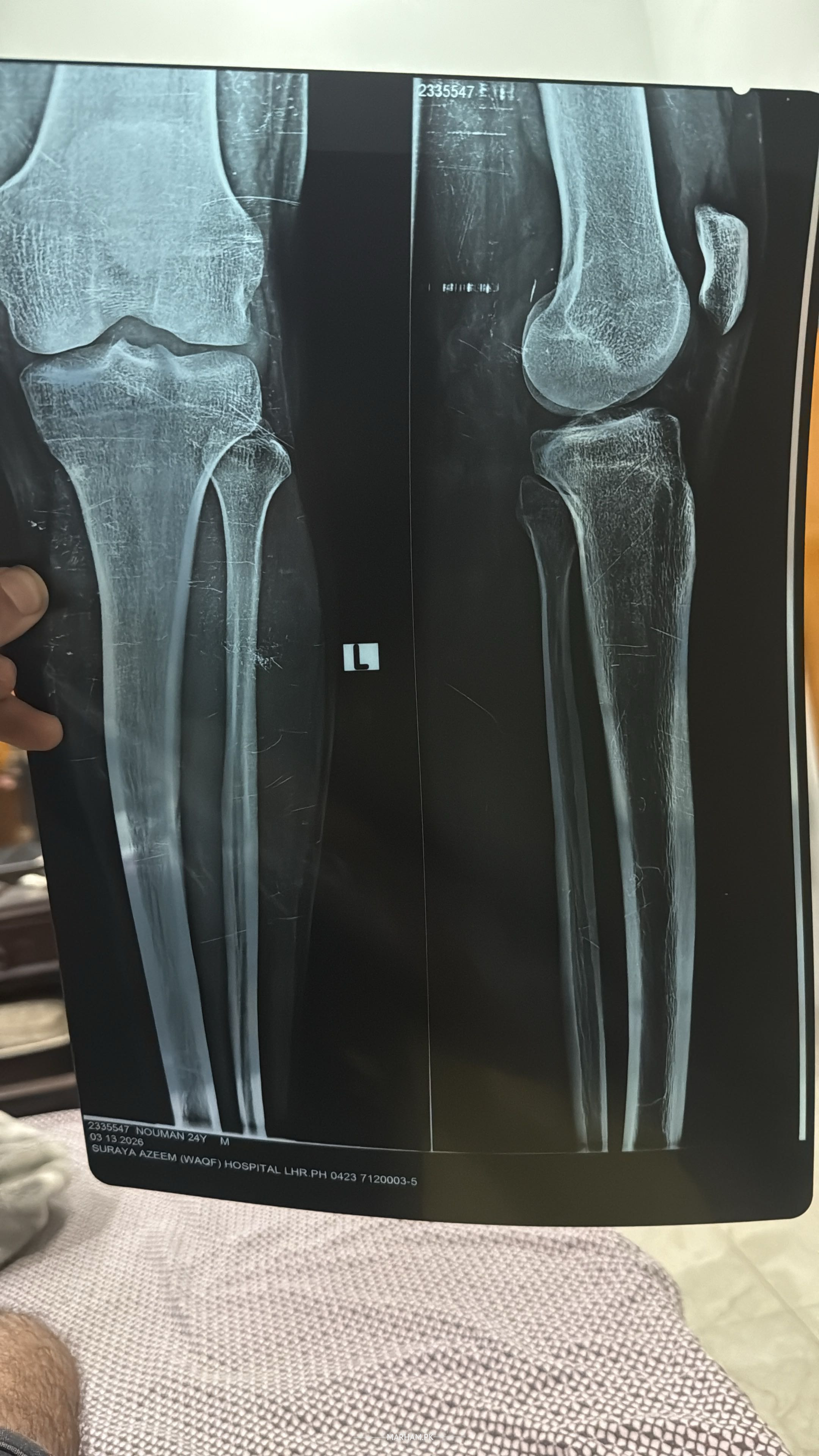
Looks one of your bone in leg Tibia is not straight. Better to see any orthopedic Surgeon near you for proper assessment and than management.
Regards
Asst. Prof. Dr. Abdul Hannan - Orthopedic Surgeon
MBBS, FACS (USA), MS (Orthopedics) | Lahore
447 Positive Reviews
Its fine… do regular excercise..
Dr. Ibtesam Haider - Orthopedic Surgeon
MBBS, FCPS Orthopaedic Surgery | Bahawalpur
35 Positive Reviews
Wa Alaikum Assalam dear, your history, examination and primary complaints matter more than any finding on the X-ray. Your primary complaint can be addressed via proper consultation to any of my competent colleagues. Whoever is convenient for you to visit, Thanks
need proper history examination to rule put cause of leg pain
I will help u through homeo medicine enshahAllha u feel batter .for proper treatment book session through marham then in detail we discuss further
The X-ray image you shared shows both knees (likely an AP and lateral view, or bilateral comparison) of what appears to be a 24-year-old male patient (based on the label: "NOUMAN 24Y M", dated around March 2025).
Key observations from the visible radiograph:
- The overall alignment of the femur, tibia, and fibula looks normal on both sides — no obvious angulation, displacement, or shortening that would suggest a fresh acute fracture of the long bones.
- The knee joint spaces appear preserved (no severe narrowing).
- No clear evidence of a complete tibial plateau fracture, distal femur fracture, or obvious patellar issues in the main views shown.
- However, there is a **small bony fragment visible near the lateral aspect of the right knee** (on the viewer's left side of the image, appearing as a separate small piece of bone just proximal/lateral to the tibial plateau/fibular head region).
This detached fragment is the most notable abnormality. It could represent one of the following (most likely in rough order of probability):
1. **Avulsion fracture** — commonly from the **fibular head** (e.g., arcuate ligament avulsion) or **lateral tibial plateau** (Segond-like fragment, though classic Segond is more anterior). Often associated with ACL injury, posterolateral corner injury, or twisting/varus stress.
2. **Chip fracture** from the proximal tibia or fibula due to direct trauma.
3. Less likely: old ununited fragment, loose body, or calcified structure (but appearance favors acute/subacute bone piece).
The rest of the bones look intact without obvious cortical breaks, step-offs, or comminution in the main shafts or articular surfaces.
You wrote "Leg is fine" — if this refers to no major long-bone fracture or gross instability, that's reasonable. However, the small avulsion/chip fragment is **not normal** and usually indicates some degree of ligamentous injury (especially posterolateral corner or ACL/PCL-related in many cases).
**Recommendation** (not medical advice — please consult the treating orthopedic doctor):
- This finding typically warrants **MRI of the knee** to evaluate ligaments (ACL, PCL, collaterals, posterolateral corner), menisci, and cartilage — X-ray alone misses soft-tissue damage.
- Clinical correlation is essential: pain location, swelling, instability, mechanism of injury (twist? fall? sports?), ability to bear weight, etc.
- If there's locking, giving way, significant swelling, or positive clinical tests (e.g., varus stress, dial test), it becomes more urgent.
If you can share more details (symptoms, how the injury happened, or doctor's initial comment), or if this X-ray is yours/your family member's, I can try to help narrow it down further. In any case, follow up with an orthopedic specialist — small fragments like this are often important clues to bigger soft-tissue problems. Take care!
it's normal
3 months ago
Dr. Ehtisham Panhwar PT - Physiotherapist
DPT , MASTER OF PHILOSOPHY (PHYSICAL THERAPY) | Karachi
121 Positive Reviews
X ray is completely fine this could be a muscular pain. Try doing some leg stretches and heat therapy. you can also use analgesic cream (rapid relief).

Get Appointment with Orthopedic Surgeon
- Best Orthopedic Surgeon in Lahore
- Best Orthopedic Surgeon in Karachi
- Best Orthopedic Surgeon in Islamabad
- Best Orthopedic Surgeon in Rawalpindi
- Best Orthopedic Surgeon in Faisalabad
- Best Orthopedic Surgeon in Peshawar
- Best Orthopedic Surgeon in Multan
- Best Orthopedic Surgeon in Quetta
- Best Orthopedic Surgeon in Gujranwala
- Best Orthopedic Surgeon in Sargodha
- View All
Diseases releated to Orthopedic Surgeon
Services releated to Orthopedic Surgeon
Find the Questions asked in Other Categories
- Questions asked from Gynecologist
- Questions asked from Dermatologist
- Questions asked from General Physician
- Questions asked from Gastroenterologist
- Questions asked from Urologist
- Questions asked from Psychiatry
- Questions asked from Pediatrician
- Questions asked from Orthopedic Surgeon
- Questions asked from Ent Specialist
- Questions asked from Dentist
68 Positive Reviews